
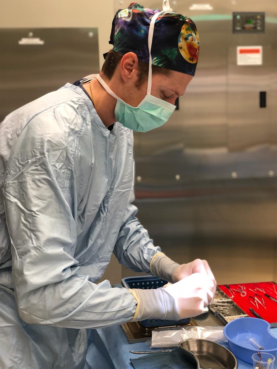
DMEK and UT-DSAEK Cornea Transplant Instructions
DMEK and UT-DSAEK SURGERY OVERVIEW
Descemet’s membrane endothelial keratoplasty (DMEK) and Ultra-Thin Descemet’s Stripping Automated Endothelial Keratoplasty (UT-DSAEK) are minimally invasive procedures used to rejuvenate the cornea when specialized endothelial pump cells stop working. These pump cells keep the cornea clear and compact, and without them, it becomes swollen, cloudy, and may develop internal droplet-like changes. DMEK and UT-DSAEK surgery are forms of cornea transplant in which human donor tissue is used to restore the clarity of the cornea. As the cornea tissue comes from human donors, it is a true gift of sight. The donor process is similar to that used for other donor organs. Serology is done on every donor and all donors are screened to exclude HIV and Hepatitis among other viruses. The donor cornea is screened carefully prior to deeming it suitable for surgical use by the eye bank and only those passing the strict parameters set by the Eye Bank Association of America are accepted for transplant.
The main difference between DMEK and UT-DSAEK is the thickness of the graft used in the surgery and the size of the wounds needed to do the surgery. The cornea graft in UT-DSAEK consists of donor endothelial cells on a membrane plus a thin layer of donor cornea stromal tissue. The cornea graft in DMEK consists only of the specialized endothelial cells on a membrane with no additional donor stromal tissue. The UT-DSAEK graft ranges from 50-100 microns thick, while a DMEK graft is only 12-15 microns thick (about the width of 1-2 red blood cells). DMEK is an advanced, modern technique involving minimally invasive incisions, often small enough that sutures are not needed to close the wounds.
As the DMEK or UT-DSAEK graft attaches to the back of the cornea, the specialized endothelial pump cells turn on and start to clear the cornea. Until they are fully pumping, an air bubble is used to hold the graft in place against the back of the cornea. As the cornea swelling clears, the vision improves. Since less tissue is transplanted in DMEK compared to DSAEK surgery, there is less risk of rejection with DMEK surgery and the quality of the vision is often better. Not all patients are candidates for DMEK surgery. Talk to your cornea specialist about which procedure may be right for you.
PREPARING FOR DMEK OR UT-DSAEK SURGERY
Try to plan ahead for the positioning requirements after surgery (lying on your back for several days). If possible, have a family member or friend assist you for the first few days. Purchase finger food or things you can eat without looking down at the plate. Prepare your sleeping arrangements so that you can sleep with your eye parallel to the ceiling (on your back). Some patients purchase periscope or mirror glasses so that they can lie in bed looking up at the ceiling but still see the TV.
If you have a history of Cardiac (Heart), Pulmonary (Lung), or other serious health conditions you will need to obtain Primary Care or Cardiology Clearance prior to surgery. This clearance will need to be faxed or sent to your surgery center in advance.
Prior to surgery, your eye surgeon may recommend a YAG laser iridotomy. This procedure is used to create a microscopic opening in your iris. The opening in the iris can help prevent angle-closure glaucoma by preventing fluid from building up the iris. Doing this laser opening prior to transplant surgery lowers your risk of developing pupillary block or angle-closure glaucoma.
Alternatives to the laser iridotomy include a surgical iridotomy or electing not to have the iridotomy. A surgical iridotomy is an opening in the iris that is made manually at the time of surgery. Electing not to have an iridotomy increases your risk of angle-closure in which the eye pressure can rise and damage your optic nerve. You could lose all vision and you could also have severe eye pain.
Prior to surgery, it is important to review all your medications with your doctor and you may be asked to adjust some medications before surgery. If you take a blood thinner, please talk to your prescribing doctor about the risks and benefits of holding it before surgery. If you are not sure if you take a blood thinner or would like more information, please talk to your primary care provider. Additional information on blood thinners around the time of surgery is provided on the home page for eye surgery instructions. You may be asked to start eye drops prior to your surgery to help reduce swelling and inflammation and risk of infection after surgery.
THE DAY OF DMEK OR UT-DSAEK SURGERY
Your surgery will be done in an outpatient surgery center or hospital and you will likely go home the same day. Continue all your usual eye drops as prescribed, including the morning of surgery. You may take your usual blood pressure or heart medications with a small sip of water in the morning unless directed otherwise by your doctor or anesthesia team.
One business day prior to your surgery someone will call you from the surgery center to discuss your arrival time for surgery. Plan to be at the surgery center for 4-6 hours.
Surgery requires FASTING. NO EATING OR DRINKING starting at MIDNIGHT the night before surgery. If your surgery is scheduled for the afternoon, then please NO EATING OR DRINKING AT LEAST 6 HOURS BEFORE your arrival time.
Please wear loose comfortable clothing. Shower and wash your hair the evening prior or morning of your surgery. WASH YOUR FACE THOROUGHLY.
No Makeup, Perfume, Lotions, or Jewelry/Valuables. No Smoking or Alcohol 24 hours prior to surgery.
You will need to arrange transportation to and from the surgery. You may NOT drive yourself.
ANESTHESIA FOR CORNEA SURGERY
ANESTHESIA FOR CORNEA SURGERY
Many patients worry if eye surgery will be painful. Modern anesthesia has advanced greatly, helping patients feel comfortable during the procedure.
In addition to local or topical anesthesia, many patients choose to have light sedation which is often administered by an anesthesiologist or nurse anesthetist during surgery.
Topical Anesthesia:
We use eye drops called proparacaine and tetracaine to numb the eye prior to surgery. Once the eye is numb, you may feel mild pressure and cold water during the surgery but not pain. It is normal to see bright lights during surgery as well.
Local Anesthesia:
In addition to topical anesthesia, your surgeon may recommend numbing the eye with an injection around the eye. This is called a peribulbar or retrobulbar block and it can help place the eye in a deeper state of numbing. This type of anesthesia is typically offered for cornea transplants or for surgeries anticipated to be longer or more challenging in nature.
Intravenous (IV) Anesthesia:
Though many patients prefer it, IV sedation is not necessary for eye surgery. Sedating medications, such as Versed and Fentanyl, are given to help relax you during your surgery. All medications are dosed incrementally to achieve the right level of sedation and comfort for each patient. Please be aware that even with IV sedation, MANY PATIENTS WILL BE AWAKE DURING SURGERY. Do not expect to be “put to sleep” as your eye surgeon often prefers that you can hold still and focus on the microscope light during the procedure.
CORNEA SURGERY POST-OPERATIVE RECOVERY
DMEK surgery is transplant surgery. Topical steroid eye drops are required to reduce the risk of graft rejection. In most cases, steroid drops are continued forever however the dose gradually is reduced to once daily several months after surgery. Stopping the steroid drop is associated with an increased risk of graft rejection, especially in the first few months after surgery. NEVER stop using your steroid eyedrop unless specifically directed to stop by your cornea transplant surgeon.
Starting immediately after surgery, you must lie flat on your back and look up to the ceiling. You will continue to lie on your back for the next 4-5 days (or until the air bubble resolves). You can take 10-minute breaks each hour to eat, use the bathroom, and do other activities of daily living. The better you are able to position, the higher your chance for successful graft attachment. Staring up at the ceiling afterward is the most important part of the surgery and the most difficult part for the patient.
It is normal for your eye to be red and to feel scratchy and sore after surgery. You may use lubricating drops to help. It normal to have very limited vision initially after surgery. As the air bubble reabsorbs you may start to notice the bubble in your vision. As the bubble clears and the cornea clears your vision will slowly improve. It is also normal to see glare and halos or foggy vision after surgery. You can expect these changes in your vision to gradually improve in the weeks following surgery.
Try to avoid looking down at the floor after surgery. When taking your 10-minute breaks and walking, you may look straight ahead or level. When you eat, try to eat finger food or things you can bring to your mouth without looking down at a plate. When you brush your teeth at night, try to spit out into a cup so you don’t need to look down. Listening to the radio or getting books on tape may help pass the time. Assuming your television is not bolted to the ceiling, you may purchase periscope or prism glasses so that you may see your TV on the wall while you are looking up at the ceiling.
Sometimes the patient can experience high eye pressure shortly after surgery which can be from angle-closure or blockage of the drain of the eye. If that occurs, you would experience significant eye pain, headache, nausea and vomiting. If you have those symptoms or if you have any concerns after surgery please call right away.
Pick up your medication eye drops PRIOR to surgery as you may be asked to start them right away after surgery. Your doctor will review your post-op medications with you so please bring them with you to your follow up appointment.
If you notice decreased vision, pain, flashes of light, changes in floaters, a curtain or veil over your vision, please call the office at 941-499-1570 right away.
RESUMING ACTIVITIES AFTER CORNEA SURGERY
RESUMING ACTIVITIES AFTER SURGERY
You may resume your normal diet and medications immediately after surgery.
Wait to drive until advised by your doctor. Sleep in the eye shield at night for 2 weeks. It is never a good idea to rub your eye, but especially avoid rubbing your eye for 1 month after surgery. You may shower after surgery but face away from the water and avoid getting water directly in your eye for a few days.
NO SWIMMING POOLS, HOT TUBS, or GARDENING (LAWN MOWING) for 3 weeks after surgery. These activities increase your risk of serious eye infection during the post-op period. Wait 3 weeks to play woodwind and brass instruments. Wait 2 months to scuba dive after eye surgery
You may wear your regular eyeglasses after surgery however the old prescription will no longer work in the surgical eye. Some people opt to remove the lens in front of the eye that had surgery. Glasses may be prescribed after surgery, though sometimes it may be advised to wait several months for the eye to heal.