Cornea Edema and Fuchs’ Corneal Dystrophy

Who was Ernst Fuchs?

Ernst Fuchs was an Austrian ophthalmologist who made many significant contributions to the field of ophthalmology. He first described the changes in Fuchs’ endothelial corneal dystrophy around 1910. His ability to characterize the disease is particularly remarkable considering the first slit lamp for eye examination was only invented in 1911 by Allvar Gullstrand.

What is Corneal Edema?

The cornea is the clear window through which the eye “sees” the world. It is the clear front part of the eye that is coated by the tear film. The cornea is made of different layers with each layer performing a different function. The surface layer and the middle layer provide barrier protection and strength to the cornea. The inner-most layer of the cornea is made of functional cells called endothelial cells. These delicate endothelial cells have a specialized function to pump water out the cornea continuously helping keep the cornea clear. Just as a boat taking on the water will sink, if the pump cells stop working the cornea takes on too much water it swells. This swelling is called corneal edema and it makes the vision will be blurry. These special endothelial pump cells live on the back surface of the cornea, inside the eye. There are many conditions in which these endothelial cells can become damaged or stop working such as aging, prior eye surgery as well as genetic conditions like Fuchs’ corneal dystrophy. Cornea edema also causes glare and poor contrast sensitivity. In severe forms, cornea edema can progress to painful blisters and put the eye at risk of infection.

What is Fuchs’ Endothelial Corneal Dystrophy?

In Fuchs’ corneal dystrophy, the specialized endothelial pump cells quit working and patients experience a gradual decline in vision and worsening of glare. The two main changes that cause the vision to decline in Fuchs’ dystrophy are corneal edema and guttae. Corneal edema is where the cornea becomes hazy and cloudy from swelling. Guttae (pronounced Goo-Tay) are “water droplet-like” changes that form on the inside of the cornea (on Descemet’s membrane) on the inner-most layer and can cause significant light scatter. Patients often describe this change as “looking through a dirty shower curtain.”

Early in the disease, the vision may clear within an hour or two after waking. In later stages, the vision will remain blurry all day. Often these symptoms progress so gradually that the patient may not notice the vision change until it is quite advanced. Fuchs’ dystrophy patients typically start to show symptoms between age 40-60 and typically progress to needing cornea transplant surgery within the following 5-10 years.

Treatment Options for Fuchs' Dystrophy

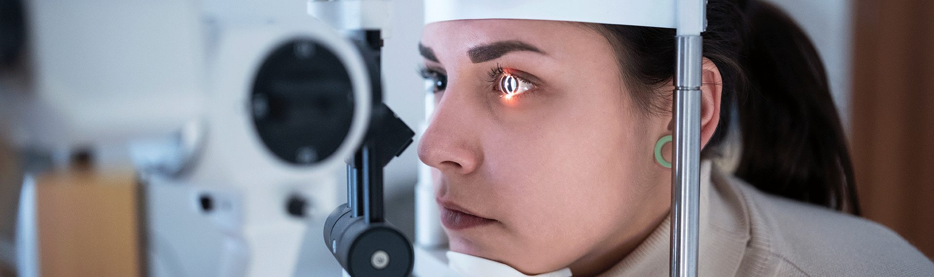
Today there are many excellent treatment options for Fuchs’ corneal dystrophy. In the early stages, medical treatment with hypertonic eye drops and ointments such as Muro 128 can be effective at reducing the swelling of the cornea. For some patients, the dystrophy will progress and the pump cells will no longer be able to keep up with the swelling. This is typically when patients will notice the blurry vision. In addition, the guttae changes can accumulate over time and can also affect the vision. It is during these later stages that patients should consider surgical options.

Surgical treatments for Fuchs’ dystrophy have progressed rapidly in the past 10 years and the visual outcomes today can be terrific. As a center of excellence for Fuchs’ dystrophy, Tailored Eyes is proud to be one of the few centers able to offer all of the treatment options for Fuchs’ dystrophy.

DSO

One option called Descemet’s Stripping Only (DSO) is where we rejuvenate the cornea without using donor cornea tissue. The central guttae changes are removed from the visual axis to clear the way and no cornea graft is inserted in the eye. This can be helpful for mild to moderate cases of Fuchs’ dystrophy. The advantage of this procedure is no donor corneal tissue is needed. The disadvantages are that the vision recovery can be slow and it may not clear the cornea in about 10% of patients. For the patients who do not regain clear vision after DSO, we would proceed with a DMEK procedure (described below) for the best vision.

DMEK

Another surgical option, Descemet’s membrane endothelial keratoplasty (DMEK), is a minimally invasive procedure used to rejuvenate the cornea. DMEK surgery is a form of cornea transplant in which human donor cornea tissue is used to restore corneal clarity. As the cornea tissue graft comes from human donors it is a true gift of sight. In DMEK, the diseased Descemet’s membrane and guttae are removed from the inside of the cornea and replaced with a donor graft of similar size and thickness. The graft is unfolded inside the eye and held in place with an air bubble. The air bubble resolves in a few days. DMEK is an advanced, modern technique in which minimally invasive incisions are made and the graft is as thin as possible. The DMEK graft contains the specialized endothelial cells on a membrane with no additional donor tissue. This means the graft is only about 15 microns thick (slightly thicker than a red blood cell) and as a result, the clarity of the cornea after surgery can be spectacular. As the DMEK graft attaches to the back of the cornea, the pump cells turn on and start to clear the cornea. As the cornea clears, the vision improves (typically over 2-4 weeks).

The advantages of DMEK are that it can provide fast vision recovery with excellent visual acuity and quality of vision. The disadvantages are that there is a positioning requirement to lie on your back looking up after surgery (so the air bubble will support the graft) and it can’t be performed in patients with glaucoma tube shunts or anterior chamber lenses. These patients can still benefit from DSAEK surgery (see below).

DSAEK

Descemet’s stripping automated endothelial keratoplasty (DSAEK), is another form of cornea transplant similar to DMEK surgery. As above, the diseased Descemet’s membrane is removed from the inside of the cornea and replaced with human donor cornea tissue. The graft is then unfolded and held in place with an air bubble. The air bubble will resolve over a few days. DSAEK is an older cornea transplant technique that involves slightly larger incisions than DMEK and the donor graft is slightly thicker. The cornea graft in DSAEK consists of donor endothelial cells plus a thin layer of donor cornea stroma (the middle layer of the cornea). As the DSAEK graft attaches to the back of the cornea, the pump cells turn on and start to clear the cornea. As the cornea clears, the vision improves. In DSAEK surgery, the vision recovery can take longer than DMEK (3-6 months), however, it is an excellent option for patients who may have difficulty lying on their back or otherwise cannot have DMEK surgery.

Why Is My Vision Blurry First Thing in the Morning?

The main reason for blurry vision in the morning is that the cornea swells overnight while you sleep. This swelling occurs because the specialized endothelial pump cells slow down at night. The endothelial pump cells require oxygen to be most active (this is called aerobic metabolism) which they typically get by diffusion through the air (when your eyelids are open). When you sleep, you close your eyelids and the oxygen levels in the cornea drop. The specialized pump cells slow down with the drop in oxygen (change to anaerobic metabolism) and the cornea swells. When you wake in the morning, the endothelial pump cells return to their active pumping state and they clear the cornea rapidly. In patients with Fuchs’ endothelial dystrophy, those pump cells are damaged and it may take extra time for them to be able to clear the cornea. In later stages of Fuchs’ dystrophy, the pump cells may not have enough remaining action to be able to clear the cornea.

Is Glare in Bright Lights and at Night Related to Fuchs' Dystrophy?

Yes. The glare is the result of light scattering as it passes through the cornea. In a healthy cornea, light passes through the cornea without significant scatter. In Fuchs’ dystrophy, the guttae (pronounced Goo-Tay) changes on the inside of the cornea cause significant light scatter. Guttae are “water droplet-like” changes that accumulate on the inside of the cornea in patients with Fuchs’ dystrophy. These changes scatter light similar to the light scattering effect of rain droplets in the atmosphere causing a rainbow.

Will My Vision Get Worse?

Fuchs’ corneal dystrophy is typically a slowly progressive condition that leads to a gradual decline in vision and worsening quality of vision. Many patients will progress to needing a surgical procedure to restore the vision within about 10 years from the time of diagnosis.

New Patient?

Fill out your intake forms now.

Fill Out Forms